
Nano-particles made of gold and silica can combine cancer treatments and imaging techniques in a single tiny sphere, researchers say.
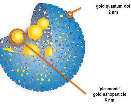 The spheres can deliver chemotherapy and photothermal therapy – and can be used for MRI imaging, near-infrared fluorescence and photo-acoustic imaging, according to the developers.
They can be activated by infrared laser to emit infrared fluorescence together with heat.
Researchers at Imperial College London and the Laboratoire de chimie de la matière condensée de Paris, France, say they have successfully been tested on human cultured cells and on laboratory mice.
They say they have solved the puzzle of how to use gold quantum dots – which have unique properties of fluorescence, magnetism and heat production compared with larger gold nanoparticles.
Until now these have been unstable but the new technique infuses them into porous silica shells and preserves their optical and magnetic properties.
The developers say this is equivalent to creating a therapeutic Swiss Army Knife with multiple functions.
The breakthrough was announced in the Proceedings of the National Academy of Sciences last week.
A spokesman said: "Tested in mice and on cultured human cells, they make it possible to combine two forms of tumour treatment and three imaging techniques. They notably have a greater drug loading and delivery capacity than carriers currently on the market, which opens interesting perspectives for cancer research."
Gold-Silica Quantum Rattles for Multimodal Imaging and Therapy. PNAS February 4, 2015
Leave a Reply