
Surgeons have safely removed brain tumour cells with the help of a chemical that creates a fluorescent marker and highlights the cancer, a conference has heard.
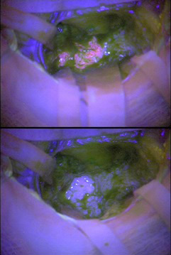 A study, presented at the 2018 National Cancer Research Institute Cancer Conference in Glasgow, UK, yesterday (4 November 2018), was undertaken with people who had suspected glioma.
The study, led by Professor Colin Watts, chair of the Birmingham brain cancer programme at the University of Birmingham, UK, used 5-aminolevulinic acid (5-ALA), which glows pink when a light is shone on it.
Kathreena Kurian, a reader/associate professor in brain tumour research at the University of Bristol and consultant neuropathologist at North Bristol NHS Trust, UK, told delegates that using the fluorescent marker helped surgeons to distinguish the most aggressive cancer cells from other brain tissue.
Dr Kurian said: "We wanted to see if using a fluorescent marker could help surgeons objectively identify high-grade tumour cells during surgery, allowing them to remove as much cancer as possible while leaving normal brain tissue intact."
The study involved 113 patients at the Royal Liverpool Hospital, Kings College Hospital in London and Addenbrooke’s Hospital, Cambridge, with suspected high-grade gliomas.
The patients, all aged between 23 and 77 years, were given a drink containing 5-ALA prior to surgery and 99 were able to be assessed for signs of fluorescence.
Surgeons used operating microscopes to help them look for fluorescent tissue while removing tumours from the patients’ brains. The tissue they removed was sent to the pathology lab where scientists could confirm the accuracy of the surgeons’ work.
Surgeons reported seeing fluorescence in 85 patients, 81 of whom were confirmed by pathologists to have high-grade disease. One was found to have low-grade disease and three could not be assessed.
No fluorescence was seen in 14 patients and only seven tumours could be subsequently evaluated by pathology. Low-grade disease was confirmed in all of them.
Professor Watts said: "Neurosurgeons need to be able to distinguish tumour tissue from other brain tissue, especially when the tumour contains fast-growing, high-grade cancer cells. This is the first prospective trial to show the benefits of using 5-ALA to improve the accuracy of diagnosing high-grade glioma during surgery. These results show that the marker is very good at indicating the presence and location of high-grade cancer cells.
"The advantage of this technique is that it may highlight more quickly high-grade disease within a tumour during neurosurgery. What this means is that more of the tumour can be removed more safely and with fewer complications, and that’s better for the patient."
Kurian K et al. Abstract number 1765: ‘Improving the intra-operative diagnosis of high-grade glioma using a fluorescence biomarker – result of the UK NCRI GALA-BIDD study.
Leave a Reply